ไฟล์:Leiomyoma of the Uterus.jpg
ไม่มีภาพที่มีรายละเอียดสูงกว่านี้
Leiomyoma_of_the_Uterus.jpg (550 × 521 พิกเซล, ขนาดไฟล์: 166 กิโลไบต์, ชนิดไมม์: image/jpeg)
| รูปภาพหรือไฟล์เสียงนี้ ต้นฉบับอยู่ที่ คอมมอนส์ รายละเอียดด้านล่าง เป็นข้อความที่แสดงผลจาก ไฟล์ต้นฉบับในคอมมอนส์
|
| คำอธิบาย |
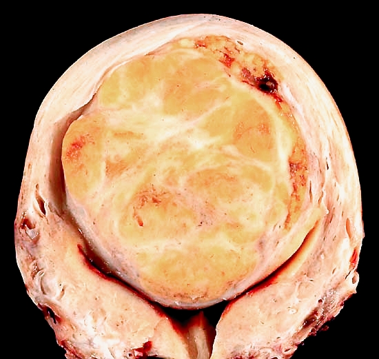
English: This hysterectomy specimen shows a large, solitary leiomyoma in the fundus, distoring the endometrial cavity into a Y shape by splaying and pressing it downwards.
Because of its unusual appearance (solitary, bright yellow, and soft), I was worried about its being a leiomyosarcoma. All of the numerous sections I took, however, showed an utterly bland neoplasm with no mitotic activity. It does make a colorful subject for the camera. The photo was taken with a Nikon FE2 on Kodak Elite daylight film, ISO 100, with blue filter to compensate for the tungsten illumination. The specimen was formalin-fixed but later soaked in 70% alcohol to return some of the native coloration. Alcohol also helps to partially dehydrate the specimen, so that, after blotting the cut surface right before taking the photo, essentially all the distracting highlights can be eliminated. |
||
| วันที่ | |||
| แหล่งที่มา | http://web2.airmail.net/uthman/specimens/index.html | ||
| ผู้สร้างสรรค์ | Ed Uthman, MD. | ||
| การอนุญาต (การใช้ไฟล์นี้ใหม่) |
|
| Camera Model | Nikon FE2 |
|---|---|
| Film speed (ISO) | 100 |
| Film | Kodak Elite daylight |
ประวัติไฟล์
คลิกวันที่/เวลาเพื่อดูไฟล์ที่ปรากฏในขณะนั้น
| วันที่/เวลา | รูปย่อ | ขนาด | ผู้ใช้ | ความเห็น | |
|---|---|---|---|---|---|
| ปัจจุบัน | 12:40, 18 มกราคม 2555 | | 550 × 521 (166 กิโลไบต์) | Hic et nunc | more natural colour |
| 05:22, 5 มิถุนายน 2549 |  | 550 × 521 (36 กิโลไบต์) | Patho | {{Information| |Description=This hysterectomy specimen shows a large, solitary leiomyoma in the fundus, distoring the endometrial cavity into a Y shape by splaying and pressing it downwards. Because of its unusual appearance (solitary, bright yellow, and |
หน้าที่มีภาพนี้
หน้าต่อไปนี้ โยงมาที่ภาพนี้:
การใช้ไฟล์ข้ามโครงการ
วิกิอื่นต่อไปนี้ใช้ไฟล์นี้:
- การใช้บน ar.wikipedia.org
- การใช้บน de.wikipedia.org
- การใช้บน de.wikibooks.org
- การใช้บน en.wikipedia.org
- การใช้บน fa.wikipedia.org
- การใช้บน hi.wikipedia.org
- การใช้บน hy.wikipedia.org
- การใช้บน id.wikipedia.org
- การใช้บน it.wikipedia.org
- การใช้บน kk.wikipedia.org
- การใช้บน la.wikibooks.org
- การใช้บน mk.wikipedia.org