ไฟล์:Human Cortical Development.png
ขนาดของตัวอย่างนี้: 520 × 600 พิกเซล ความละเอียดอื่น: 208 × 240 พิกเซล | 416 × 480 พิกเซล | 666 × 768 พิกเซล | 888 × 1,024 พิกเซล | 2,303 × 2,656 พิกเซล
ดูภาพที่มีความละเอียดสูงกว่า (2,303 × 2,656 พิกเซล, ขนาดไฟล์: 1,022 กิโลไบต์, ชนิดไมม์: image/png)
| รูปภาพหรือไฟล์เสียงนี้ ต้นฉบับอยู่ที่ คอมมอนส์ รายละเอียดด้านล่าง เป็นข้อความที่แสดงผลจาก ไฟล์ต้นฉบับในคอมมอนส์
|
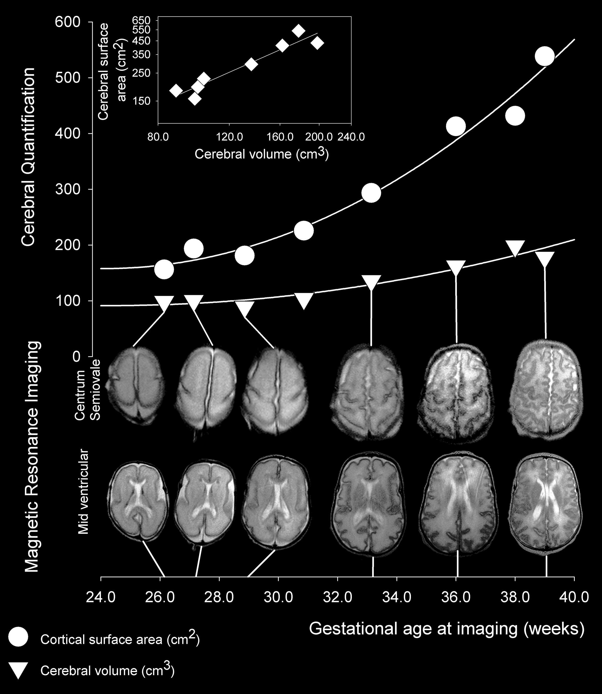
| คำอธิบาย | The images show slices through the brain at the mid-ventricular level and at the level of the centrum semiovale from six of the eight MR images obtained between 26 and 39 week gestational age; images obtained at 30 and 38 weeks are omitted for graphical clarity. Measured values for cerebral volume (triangles) and cortical surface area (circles) are related to relevant image pairs by straight lines. The insert displays a scatter plot in log-log coordinates of cortical surface area and cerebral volume (diamonds), showing a linear relationship that indicates power law scaling of cortical surface area relative to cerebral volume in this individual. | ||
| วันที่ | |||
| แหล่งที่มา | Kapellou O, Counsell SJ, Kennea N, Dyet L, Saeed N, et al. (2006) Abnormal Cortical Development after Premature Birth Shown by Altered Allometric Scaling of Brain Growth. PLoS Med 3(8): e265. doi:10.1371/journal.pmed.0030265 | ||
| ผู้สร้างสรรค์ | Kapellou O, Counsell SJ, Kennea N, Dyet L, Saeed N, et al. | ||
| การอนุญาต (การใช้ไฟล์นี้ใหม่) |
|
ประวัติไฟล์
คลิกวันที่/เวลาเพื่อดูไฟล์ที่ปรากฏในขณะนั้น
| วันที่/เวลา | รูปย่อ | ขนาด | ผู้ใช้ | ความเห็น | |
|---|---|---|---|---|---|
| ปัจจุบัน | 22:13, 5 มกราคม 2553 | | 2,303 × 2,656 (1,022 กิโลไบต์) | Was a bee | {{Information |Description=The images show slices through the brain at the mid-ventricular level and at the level of the centrum semiovale from six of the eight MR images obtained between 26 and 39 week gestational age; images obtained at 30 and 38 weeks |
หน้าที่มีภาพนี้
หน้าต่อไปนี้ โยงมาที่ภาพนี้:
การใช้ไฟล์ข้ามโครงการ
วิกิอื่นต่อไปนี้ใช้ไฟล์นี้:
- การใช้บน ar.wikipedia.org
- การใช้บน de.wikipedia.org
- การใช้บน en.wikipedia.org
- การใช้บน es.wikipedia.org
- การใช้บน outreach.wikimedia.org
- การใช้บน pt.wikipedia.org
- การใช้บน uk.wikipedia.org