ไฟล์:Echinococcus Life Cycle.svg

ขนาดของตัวอย่าง PNG นี้ของไฟล์ SVG นี้: 629 × 600 พิกเซล ความละเอียดอื่น: 252 × 240 พิกเซล | 504 × 480 พิกเซล | 806 × 768 พิกเซล | 1,074 × 1,024 พิกเซล | 2,149 × 2,048 พิกเซล | 1,280 × 1,220 พิกเซล
ดูภาพที่มีความละเอียดสูงกว่า ((ไฟล์ SVG, 1,280 × 1,220 พิกเซล, ขนาดไฟล์: 643 กิโลไบต์))
| รูปภาพหรือไฟล์เสียงนี้ ต้นฉบับอยู่ที่ คอมมอนส์ รายละเอียดด้านล่าง เป็นข้อความที่แสดงผลจาก ไฟล์ต้นฉบับในคอมมอนส์
|
ความย่อ
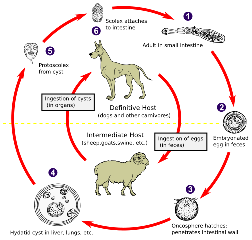
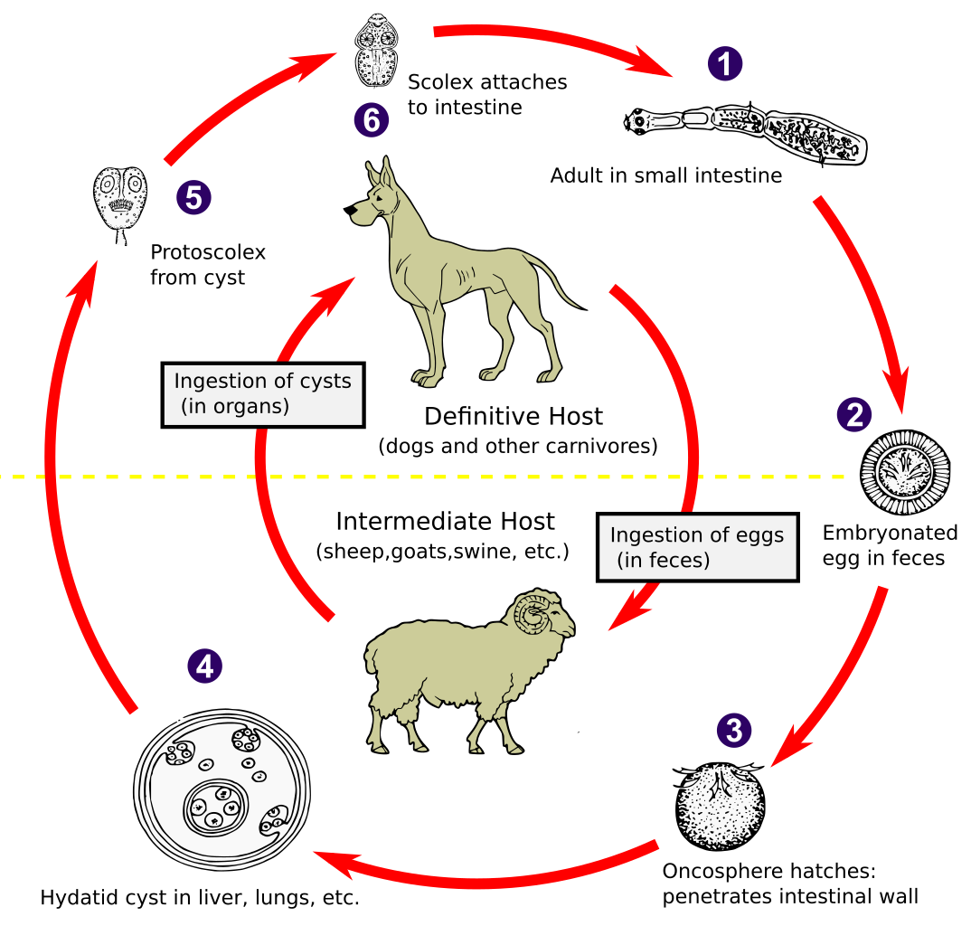
| คำอธิบาย | The adult Echinococcus granulosus (3 to 6 mm long) [1] resides in the small bowel of the definitive hosts (dogs or other carnivores). Gravid proglottids release eggs [2] that are passed in the feces. After ingestion by a suitable intermediate host (under natural conditions: sheep, goat, swine, cattle, horses, camel), the egg hatches in the small bowel and releases an oncosphere [3] that penetrates the intestinal wall and migrates through the circulatory system into various organs, especially the liver and lungs. In these organs, the oncosphere develops into a cyst [4] that enlarges gradually, producing protoscolices and daughter cysts that fill the cyst interior. The definitive host becomes infected by ingesting the cyst-containing organs of the infected intermediate host. After ingestion, the protoscolices [5] evaginate, attach to the intestinal mucosa [6] and develop into adult stages [1] in 32 to 80 days. The same life cycle occurs with E. multilocularis (1.2 to 3.7 mm), with the following differences: the definitive hosts are foxes, and to a lesser extent dogs, cats, coyotes and wolves; the intermediate host are small rodents; and larval growth (in the liver) remains indefinitely in the proliferative stage, resulting in invasion of the surrounding tissues. With E. vogeli (up to 5.6 mm long), the definitive hosts are bush dogs and dogs; the intermediate hosts are rodents; and the larval stage (in the liver, lungs and other organs) develops both externally and internally, resulting in multiple vesicles. E. oligarthrus (up to 2.9 mm long) has a life cycle that involves wild felids as definitive hosts and rodents as intermediate hosts. Humans become infected by ingesting eggs , with resulting release of oncospheres in the intestine and the development of cysts in various organs. Image adapted from original available at the United States Centres for Disease Control Parasitology Identification Laboratory ([1]). |
| วันที่ | |
| แหล่งที่มา |
This file was derived from: Echinococcus Life Cycle.png: |
| ผู้สร้างสรรค์ |
CDC เวกเตอร์: 🎱 |
| SVG genesis | ไฟล์ภาพกราฟิกส์เวกเตอร์ W3C ตรวจสอบแล้วมีข้อผิดพลาดในไฟล์นี้ สร้างขึ้นโดยใช้ Other tools |
การอนุญาตใช้สิทธิ
This image is a work of the Centers for Disease Control and Prevention, part of the United States Department of Health and Human Services, taken or made as part of an employee's official duties. As a work of the U.S. federal government, the image is in the public domain.
|
บันทึกการอัพโหลด
This image is a derivative work of the following images:
- Echinococcus Life Cycle.png licensed with PD-USGov-HHS-CDC
- 2007-01-24T10:54:56Z Pngbot 600x571 (45555 Bytes) optimized with optipng
- 2005-04-26T01:48:50Z FirstPrinciples~commonswiki 600x571 (55999 Bytes) Smaller & clearer
- 2005-04-26T01:36:23Z FirstPrinciples~commonswiki 800x761 (80990 Bytes)
Uploaded with derivativeFX
ประวัติไฟล์
คลิกวันที่/เวลาเพื่อดูไฟล์ที่ปรากฏในขณะนั้น
| วันที่/เวลา | รูปย่อ | ขนาด | ผู้ใช้ | ความเห็น | |
|---|---|---|---|---|---|
| ปัจจุบัน | 08:31, 1 กุมภาพันธ์ 2564 | | 1,280 × 1,220 (643 กิโลไบต์) | Pixelsquid | Resized. |
| 03:44, 1 กุมภาพันธ์ 2564 |  | 320 × 305 (460 กิโลไบต์) | Pixelsquid | == {{int:filedesc}} == {{Information |Description=The adult Echinococcus granulosus (3 to 6 mm long) [1] resides in the small bowel of the definitive hosts (dogs or other carnivores). Gravid proglottids release eggs [2] that are passed in the feces. After ingestion by a suitable intermediate host (under natural conditions: sheep, goat, swine, cattle, horses, camel), the egg hatches in the small bowel and releases an oncosphere [3] that penetrates the intestinal wall and migrates through the... |
หน้าที่มีภาพนี้
หน้าต่อไปนี้ โยงมาที่ภาพนี้:
การใช้ไฟล์ข้ามโครงการ
วิกิอื่นต่อไปนี้ใช้ไฟล์นี้:
- การใช้บน ar.wikipedia.org
- การใช้บน arz.wikipedia.org
- การใช้บน be.wikipedia.org
- การใช้บน bs.wikipedia.org
- การใช้บน ca.wikipedia.org
- การใช้บน dag.wikipedia.org
- การใช้บน el.wikipedia.org
- การใช้บน en.wikipedia.org
- การใช้บน es.wikipedia.org
- การใช้บน fa.wikipedia.org
- การใช้บน ga.wikipedia.org
- การใช้บน gl.wikipedia.org
- การใช้บน hi.wikipedia.org
- การใช้บน hu.wikipedia.org
- การใช้บน hy.wikipedia.org
- การใช้บน ia.wikipedia.org
- การใช้บน id.wikipedia.org
- การใช้บน is.wikipedia.org
- การใช้บน it.wikipedia.org
- การใช้บน ja.wikipedia.org
- การใช้บน ko.wikipedia.org
- การใช้บน ky.wikipedia.org
- การใช้บน lt.wikipedia.org
- การใช้บน mk.wikipedia.org
- การใช้บน ml.wikipedia.org
- การใช้บน ms.wikipedia.org
- การใช้บน nl.wikipedia.org
- การใช้บน om.wikipedia.org
- การใช้บน or.wikipedia.org
- การใช้บน pl.wikipedia.org
- การใช้บน pt.wikipedia.org
- การใช้บน ro.wikipedia.org
- การใช้บน ru.wikipedia.org
- การใช้บน sl.wikipedia.org
- การใช้บน sr.wikipedia.org
- การใช้บน sv.wikipedia.org
- การใช้บน tl.wikipedia.org
- การใช้บน tr.wikipedia.org
- การใช้บน uk.wikipedia.org
- การใช้บน uz.wikipedia.org
- การใช้บน vi.wikipedia.org
- การใช้บน www.wikidata.org
ดูการใช้ข้ามโครงการเพิ่มเติมของไฟล์นี้